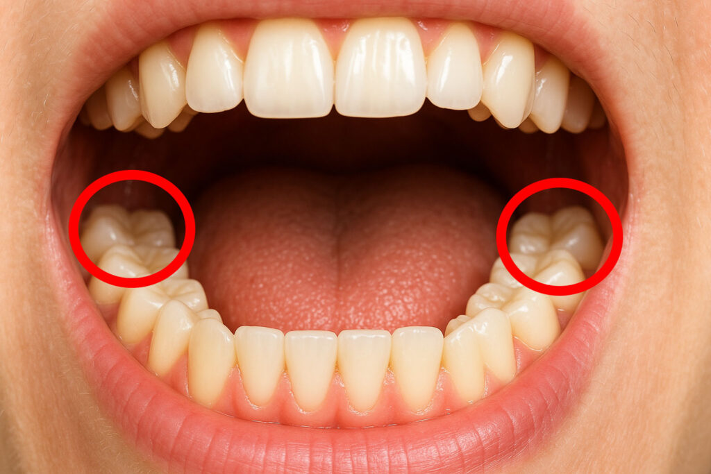
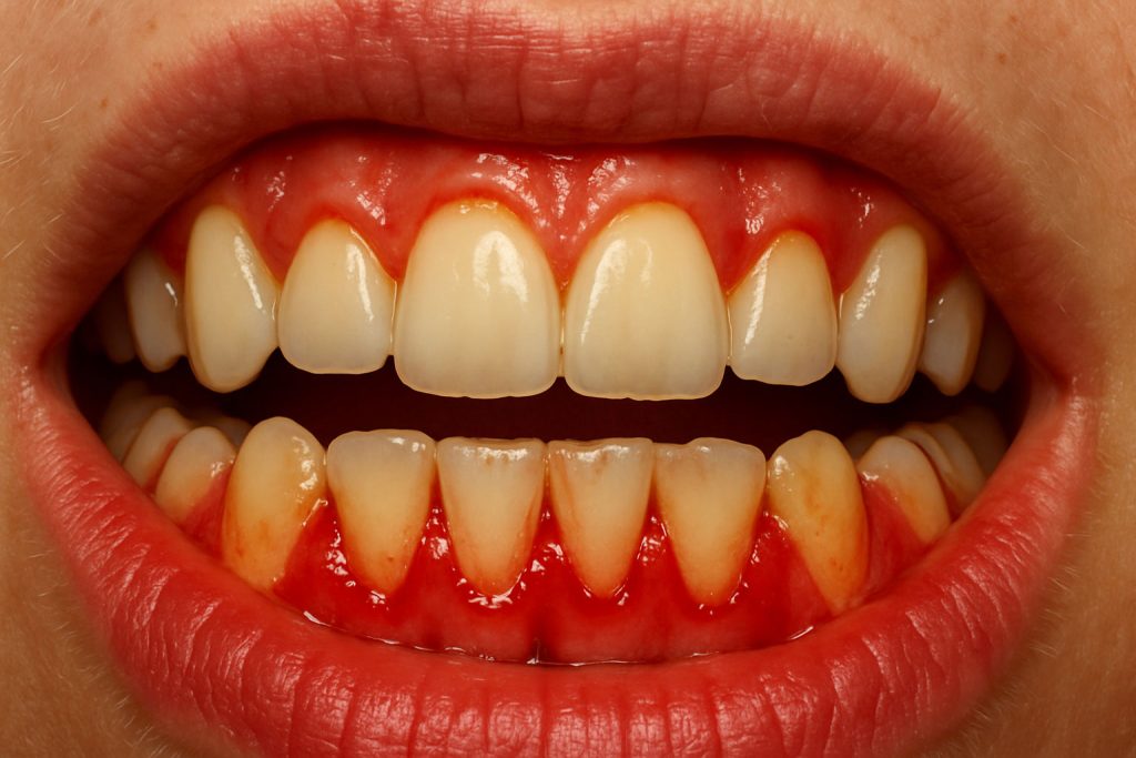
Dental neuralgia manifests itself as sharp pains or intense electric shocks in the teeth, jaw, or facial area, which can appear suddenly and last only a few seconds, but reappear very frequently. In this article, we will explain what dental neuralgia is, why it occurs, and how to distinguish it from other types of orofacial pain.
Our goal is to provide you with clear information about its causes, diagnosis, and treatments—both immediate and long-term—so that you can relieve the pain and regain your quality of life.
Whether you suffer from episodes of neuralgia or want to know how to help someone who does, here you will find a practical and accessible guide that will walk you through the process step by step.
>>> Do you live in Mallorca? Book your free first appointment <<<
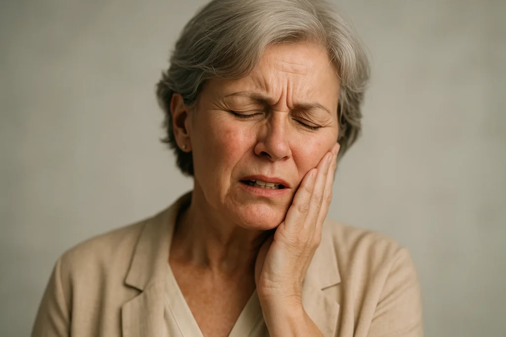
What is dental neuralgia?
Dental neuralgia is a type of neuropathic pain that affects the nerves that innervate the teeth and surrounding tissues. It is characterized by episodes of sharp, intense, short-lived pain, often described as “electric shocks” or “cramps,” which are triggered by stimuli as mild as toothbrush contact, cold air, or even the simple act of speaking.
Unlike the discomfort caused by tooth decay or pulp inflammation, the origin of neuralgia is not in the tooth itself, but in an alteration of the trigeminal nerve—or one of its branches—that causes neuronal hyperexcitation and a disproportionate pain response.
Incidence of dental neuralgia in the general population
It is estimated that dental neuralgia affects between 2% and 5% of the population throughout their lifetime, although most cases correspond to trigeminal neuralgia with pain radiating to the dental area. The annual incidence ranges from 4 to 13 new cases per 100,000 inhabitants, with a slight predominance in women (1.5–2 times more than in men). The risk increases with age, being more common after the age of 50, and peaks between the ages of 60 and 70. Although rare in people under 30, there are cases associated with secondary causes (multiple sclerosis, tumors) that can appear in young adults. Knowing these figures is vital to differentiate neuropathic pain from other more common dental conditions, so that a specialized evaluation is requested as soon as the first episodes of electric shocks or paroxysmal pain appear.
The importance of addressing neuropathic pain
Orofacial neuropathic pain can limit eating and phonation, making everyday activities such as talking or eating normally difficult. In addition, repeated attacks interrupt nighttime rest, worsening sleep quality and increasing daytime fatigue. Without proper treatment, episodes of pain tend to become more frequent and longer lasting, promoting chronicity and the development of nervous hyperreactivity. This prolonged persistence increases anxiety and stress, which in turn can intensify the perception of pain.
Addressing dental neuralgia early allows for the selection of specific therapies—pharmacological or physical—to modulate neuronal hyperexcitation. An accurate professional diagnosis reduces the risk of unnecessary or ineffective treatments and significantly improves well-being and pain responsiveness.
What causes dental neuralgia?
Dental neuralgia arises from a dysfunction in the transmission of nerve impulses from the trigeminal nerve, which is responsible for facial sensitivity.
- Peripheral vs. central neuropathic origin:
- Peripheral: Damage or compression of the nerve along its path (e.g., by pulsating arteries, tumors, or scar tissue after dental surgery).
- Central: Alterations in the nerve pathways of the brainstem, such as in multiple sclerosis, which affect the conduction of pain signals.
- Factors that aggravate neuralgia:
- Sudden changes in temperature (cold air, very hot drinks).
- Light mechanical pressure (gentle brushing, chewing soft foods).
- Stress and fatigue, which can increase the excitability of neurons.
- Main triggers and types of neuralgia:
- Classic trigeminal neuralgia: No apparent cause beyond vascular compression.
- Secondary neuralgia: Associated with systemic diseases (multiple sclerosis, brain tumors) or sequelae of facial surgery.
- Painful paroxysms: Brief, intense, and recurrent episodes, separated by periods of remission.
Understanding these mechanisms allows for a more accurate diagnosis and selection of the most appropriate treatment, whether pharmacological, surgical, or through neuromodulation techniques.
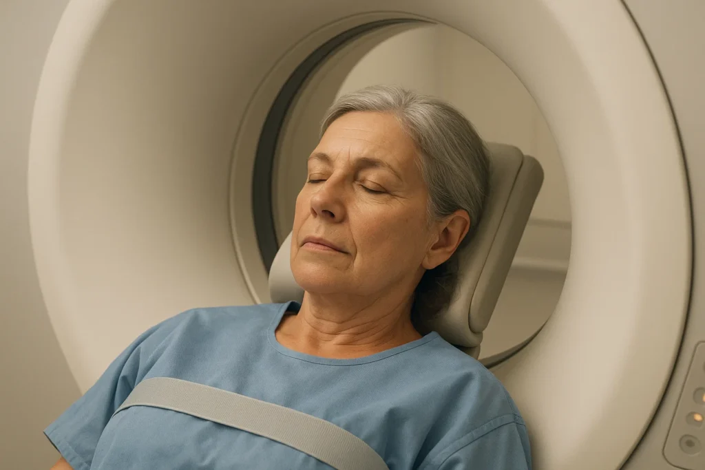
Diagnosis of dental neuralgia
The diagnosis is based on the patient’s medical history and physical examination:
- Description of the pain: The patient describes brief, intense, stabbing or “electric” episodes that last for seconds and recur in the same area.
- Specific triggers: Identify stimuli that cause pain (brushing, air, chewing, talking).
- Neurological examination: Assess facial sensitivity with thermal and tactile stimuli, ruling out loss of function or motor deficit.
- Imaging tests:
- Magnetic resonance imaging (MRI): To detect central causes (multiple sclerosis, tumors) or vascular compression of the trigeminal nerve.
- Differential diagnosis:
- Pain from caries, pulpitis, or abscesses (more continuous and reactive to thermal pulses).
- Myofascial or temporomandibular joint pain (more diffuse and associated with jaw movements).
- Cluster headache or migraine (different location and characteristics).
An accurate and early diagnosis allows pharmacological treatment or minimally invasive interventions to be started before the pain becomes chronic and more difficult to control.
Rapid resolution and initial management
Local anesthetic block
In crisis situations, nerve block with local anesthetic offers almost instant relief. By injecting lidocaine or bupivacaine into the affected trigeminal nerve branch, the transmission of pain impulses is temporarily interrupted. This brief, outpatient procedure allows the diagnosis to be confirmed and drastically reduces the intensity of pain attacks for several hours, facilitating the initiation of longer-term treatment.
First-line anticonvulsants
At the same time, anticonvulsants are the cornerstone of initial treatment. Medications such as carbamazepine or oxcarbazepine act by stabilizing the neuronal membrane and reducing nerve hyperexcitability.
They are started at low doses to assess tolerance and are gradually increased until the therapeutic threshold is reached. Many patients notice a significant reduction in both the frequency and intensity of electrical discharges after a few days of treatment.
NSAIDs and mild analgesics
Although NSAIDs and conventional analgesics are not always sufficient for pure neuropathic pain, they can offer support in the early stages or during dose adjustments of anticonvulsants. Their use should be occasional and under medical supervision to avoid gastrointestinal or renal side effects.
Cold compresses or gentle local heat
As non-pharmacological measures, cold compresses applied to the painful area temporarily relieve the stabbing sensation by reducing inflammation and nerve conduction velocity. In some cases, alternating cold and moderate heat (warm towels) helps break the pain cycle.
Avoiding triggers
Finally, it is essential to identify and avoid the triggers that cause the paroxysms. Brushing with a soft-bristled toothbrush, chewing moderately, and protecting the face from cold drafts or sudden changes in temperature reduces the occurrence of new episodes while the treatment begins to take effect.
Dental neuralgia treatments and therapeutic approaches
Maintenance pharmacotherapy
Anticonvulsants: After the initial phase with carbamazepine or oxcarbazepine, switching to gabapentin or pregabalin may be considered if tolerance is poor or side effects arise. These drugs modulate calcium channels in neurons, reducing the release of excitatory neurotransmitters and attenuating trigeminal hyperexcitability.
Tricyclic antidepressants: Amitriptyline or nortriptyline, in low doses, can help in resistant cases, acting on the serotonergic and noradrenergic systems, which increases the pain threshold.
Minimally invasive interventions
Percutaneous glycerol or phenol blocks: Fluoroscopically or ultrasound-guided injection into Gasser’s ganglion (trigeminal) to selectively destroy painful fibers. Provides prolonged relief (months to years) but with risk of residual numbness.
Pulsed radiofrequency or thermocoagulation: Application of controlled thermal energy to the nerve root to deactivate Aδ and C fibers, with the aim of relieving pain without causing permanent hypoesthesia.
Microvascular decompressive surgery
Indicated in classic trigeminal neuralgia with vascular compression confirmed by MRI. It consists of displacing the pinching artery of the nerve and interposing an interpositional material (Teflon fabric) to avoid pulsatile contact. It offers relief rates of 70-90% in the long term, although with a risk of cranial complications.
Complementary and supportive therapies
Transcutaneous electrical nerve stimulation (TENS): Electrodes placed in the trigeminal territory offer modulations of the pain gate (gate control theory), reducing paroxysmal episodes.
Acupuncture: In some studies it provides symptomatic improvement, possibly through the release of endorphins and pain modulators.
Biofeedback and relaxation techniques: Strategies to reduce muscle tension and stress, factors that can precipitate or aggravate flare-ups.
Rehabilitation and multidisciplinary support
Facial physiotherapy: Gentle massage and relaxation exercises of the masticatory musculature decrease muscle coactivation and relieve pressure on the nerve.
Psychology: Chronic pain management, stress coping and anxiety control strategies, key to reduce the frequency of attacks in chronic neuralgia.
A staged and personalized approach, combining these options according to each patient’s response and tolerance, maximizes the chances of pain control and improves quality of life.
| Characteristic | Dental neuralgia | Trigeminal neuralgia |
|---|---|---|
| Affected area | Teeth and surrounding tissues (distal branches) | Any of the three branches of the trigeminal nerve (V₁, V₂, V₃) |
| Type of pain | Sharp, brief, triggered by oral stimuli (brushing, air, cold) | Intense, like "electric shocks," along the face (eye, cheek, jaw) |
| Origin | Peripheral alteration of intraoral trigeminal fibers | Vascular compression at the nerve root, multiple sclerosis, or other central causes |
| Diagnosis | Rule out caries/dental inflammation; alveolar blocks | Neurological evaluation, MRI, and trigeminal provocation tests |
| Initial treatment | Anticonvulsants + local alveolar branch blocks | Carbamazepine/pharmacotherapy + percutaneous trigeminal block |
| Advanced interventions | Definitive alveolar branch blocks | Microvascular decompression, rhizotomy, or radiofrequency |
| Prognosis | Good response to dental treatment and local blocks | Variable; may require microvascular surgery for prolonged relief |
Preventive measures and daily care
To minimize the recurrence of episodes and improve your quality of life, it is essential to incorporate the following preventive measures and daily care:
- Stress management and relaxation habits
- Practice deep breathing exercises, meditation, or progressive relaxation techniques daily to reduce facial muscle tension and neural overexcitement.
- Spend at least 10–15 minutes a day on activities that inspire calm (gentle yoga, short walks, reading).
- Protection from physical triggers
- Avoid direct contact with cold drafts: wear a scarf or light shawl when you go outside and keep the indoor environment warm. Use neutral-flavored toothpaste to avoid irritating the nerves.
- Proper oral hygiene and daily routine
- Brush your teeth at least twice a day, paying special attention to the root areas where neuralgia often occurs.
- Complete your hygiene routine with dental floss or soft interdental brushes, and alcohol-free mouthwashes that do not increase nerve sensitivity.
- Modulation of diet and food temperature
- Eat food at room temperature: avoid very cold or very hot foods and drinks that can trigger pain.
- Limit your consumption of highly acidic citrus fruits, hot spices, and carbonated beverages that can irritate nerve endings.
- Sleeping position and care
- Sleep with your head slightly elevated to reduce possible vascular pressure on the trigeminal nerve.
- Use ergonomic pillows that keep the cervical spine aligned, reducing tension in the facial muscles.
- Regular visits to the dentist and medical follow-up
- Schedule checkups every 6–12 months to detect any dental, joint, or muscle problems that could aggravate the neuralgia.
- Always inform your dentist or neurologist of any changes in the frequency, intensity, or characteristics of the pain so that the treatment plan can be adjusted.
- Maintenance medication support
- Continue with the prophylactic medication prescribed by your doctor (anticonvulsants, antidepressants) and do not stop treatment without consulting your doctor.
- Keep a small daily record of your pain and possible triggers (food, activities, stress) to share at your follow-up appointments.
Consistently implementing these practices not only helps prevent new outbreaks, but also improves overall well-being and facilitates the effectiveness of medical treatments.

When to see a professional
Persistent pain despite initial treatment: If the episodes of pain do not subside after applying the anesthetic block protocol and starting medication, it is necessary to reevaluate the diagnosis and treatment plan.
Increased frequency or intensity of flare-ups: An increase in the frequency of flare-ups or their increasing intensity indicates that drug therapy or preventive measures need to be adjusted.
Intolerable side effects: If the medication causes nausea, excessive drowsiness, dizziness, or other adverse effects that affect your quality of life, consult your doctor to evaluate alternatives or changes in medication.
Signs of neurological complications: Tingling, loss of sensation in the face, double vision, or muscle weakness in the facial region are warning signs that require urgent attention.
Questions about the use of devices or techniques: If you have difficulty using compresses, TENS, or any other recommended device, or if you experience discomfort with the nerve block, ask for help to correct the technique.
Need for advanced interventions: When definitive percutaneous blocks, radiofrequency, or microvascular surgery need to be considered, the dentist or neurologist will refer you to a specialized center to plan the procedure.
Impact on your daily life: If pain significantly interferes with your rest, eating, communication, or mood, multidisciplinary follow-up (neurology, dentistry, psychology) is key to regaining your well-being.
In any of these cases, do not delay your visit: early and appropriate management prevents complications, reduces the risk of chronic pain, and improves your quality of life.
Final summary
Dental neuralgia is an intense and disabling disorder, but in most cases it can be controlled with a stepwise approach combining medication, nerve blocks, and, if necessary, minimally invasive techniques or microvascular surgery. Successful management requires an accurate diagnosis, aggressive initial treatment to stop the painful discharges, and then maintenance therapy tailored to each patient, including anticonvulsants or antidepressants and measures to protect against physical and emotional triggers. Complementing pharmacotherapy with facial physical therapy, relaxation techniques, and psychological support improves long-term outcomes. Regular visits to the dentist and neurologist, as well as monitoring oral hygiene habits and stress management, are essential to reduce recurrence and preserve quality of life.
FAQs about dental neuralgia
How long does drug treatment last?
The acute phase of neuralgia is treated for several weeks or months until the pain is under control. After that, many patients continue to take lower doses of anticonvulsants or antidepressants as long-term maintenance therapy, always under medical supervision.
Can dental neuralgia affect children?
It is rare in childhood, but it can occur in adolescents with a history of trauma or malocclusion. In these cases, the therapeutic approach is adapted to their age and weight, prioritizing non-invasive techniques and adjusted doses.
How long does it take to see improvement?
With proper treatment, many patients experience relief from flare-ups within a few days. Almost complete elimination of pain may require 2 to 4 weeks of medication adjustment and complementary measures.
What should you do if flare-ups recur despite treatment?
If flare-ups persist or return quickly, you should consult your doctor again. It may be necessary to optimize the dosage, change the medication, consider a permanent nerve block, or consider microvascular surgery.
Are there any useful home remedies?
Techniques such as applying gentle local heat, chewing sugar-free gum to stimulate saliva, or performing muscle relaxation exercises can complement treatment, but they are never a substitute for medical therapy and guided nerve blocks.
 Whatsapp


